
RL environments built from clinician reads.
A radiologist or pathologist reads the case. The read becomes a task. An agent works it in a real viewer and is scored against the clinician.
RAD-T-0142Slice142 / 310Radiologist reading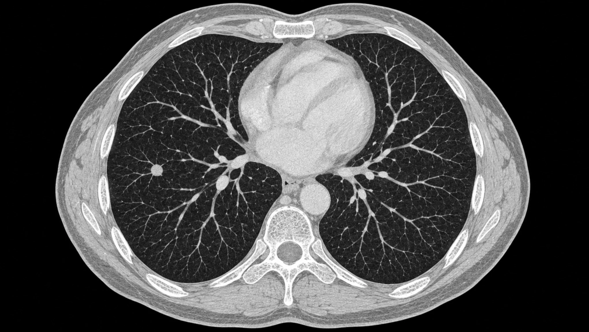
PATH-T-0031Magnification2.5×Pathologist readingWhat makes them different.
- 01
Every task starts as a clinician’s read.
A radiologist or pathologist reads the case, a second clinician checks it, and only approved reads become tasks.
- 02
Graders check clinical facts.
Each check compares one thing to the clinician’s read: where the finding is, its size, its grade. No model decides the score.
- 03
Shortcuts score nothing.
A report without evidence behind it, a guess that fits every case, a mark in the wrong place: each one earns no reward.
How an episode runs.
One episode is one case. The agent acts, the environment answers with what the viewer shows, and one reward comes back at the end, scored against a read the agent never sees.
Environment
Fixed during trainingCase
One per episodeChest CT. Find each nodule, outline it and describe how it looks.
Viewer
What the agent can do- Scroll
- Window
- Zoom
- Outline
- Measure
- Report
Each action returns the new view as an image, the way the viewer would show it to a clinician.
Held back
In no observation- Location
- slice 142
- Long axis
- 8 mm
- Texture
- solid
Approved by a second radiologist before it becomes a task.
RL training
Chest CT. Find each nodule, outline it and describe how it looks.
Scrolls the stack and stops on the nodule.
ObservationSlice 142 · lung windowOutlines it and measures the long axis.
ObservationOutline on slice 142 · long axis 7 mmPart-solid nodule in the right lung, slice 142.
Long axis 7 mm.
0.67- Location
- Long axis
- Texture
Where frontier models break.
They handle the software. They miss the medicine. That gap is what these environments train.
Radiology
They can drive the viewer. They miss what is in it.
Nine in ten runs work the imaging tools correctly. One in four finds the abnormality without guidance, and one in three places it correctly in 3D.
Frontier models on our tasks, scored by the same graders as the agents you train.
Built for radiology and pathology.
Each specialty gets its own environment, with its own cases, viewer and graders.
| Radiology | Pathology | |
|---|---|---|
| The agent | The agent reads imaging studies the way a radiologist does: it scrolls, windows and measures, then reports what it finds. | The agent reads slides the way a pathologist does: it pans and zooms across the tissue, marks what it finds and reports it. |
| Works on | Imaging studies, slice by slice | Whole-slide images |
| Tools | Scroll, window, zoom, outline, measure | Pan, zoom, outline, measure |
| Hands in | Marks on the study and a written report | Marks on the slide and a written report |
| Graded by | Deterministic verifiers | Deterministic verifiers |
| Why it is hard | The search runs through depth. On one slice a small nodule looks like a vessel; it only shows itself when followed through the stack. | The search runs across area. Cancer can be a few crowded glands on a gigapixel slide, and the grade follows the worst pattern anywhere on it. |